- Call Anytime: (414) 219-9990
- info@mypetsurgery.com
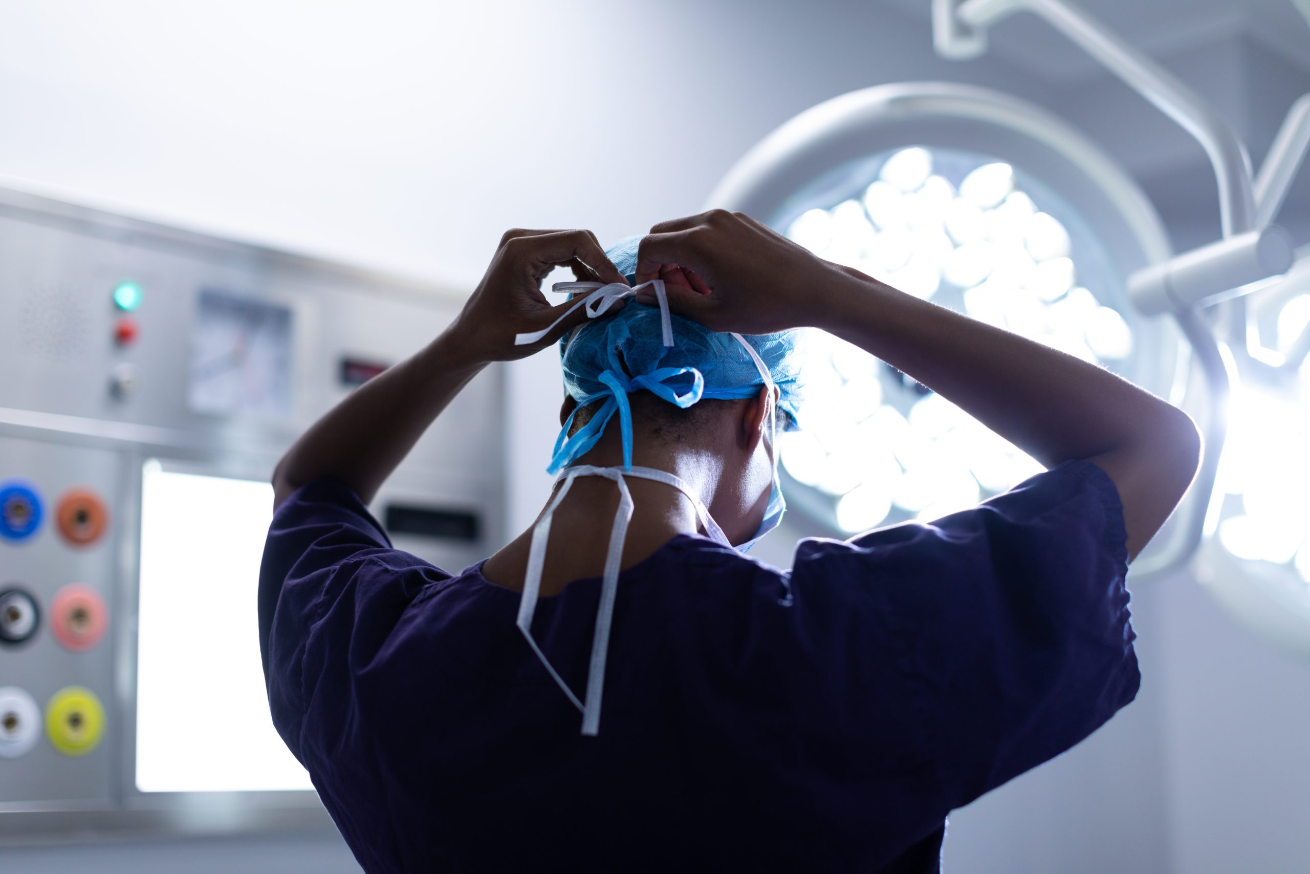
Tracheal Collapse
What is Tracheal Collapse?
Tracheal collapse is a chronic, progressive, irreversible disease affecting the trachea (windpipe) and lower airways. The trachea consists of flexible cartilage rings that help keep the airway open during breathing, movement, and coughing. However, in some dogs, these cartilage rings weaken and flatten out over time, leading to tracheal collapse. The condition most commonly affects small breed dogs, including Yorkshire terriers, Pomeranians, Poodles, and Chihuahuas.
Indications:
- Upper Airway Obstruction:
- Tracheal collapse is indicated when upper airway obstruction poses a life-threatening risk.
- It is especially relevant when the orotracheal route (through the mouth) is unavailable.
- Long-Term Positive Pressure Ventilation (PPV):
- In critically ill patients requiring mechanical ventilation, tracheostomy allows direct access to the trachea.
- Oral or Pharyngeal Surgery:
- When orotracheal intubation is not feasible (e.g., due to mouth closure), tracheostomy provides an alternative route for ventilation.
- Preoperative Management:
- Prior to definitive treatment for upper airway obstructions (e.g., allowing referral or processing biopsy specimens).
- For transient soft tissue obstructions (swelling after upper respiratory tract surgery, trauma, or inflammation).
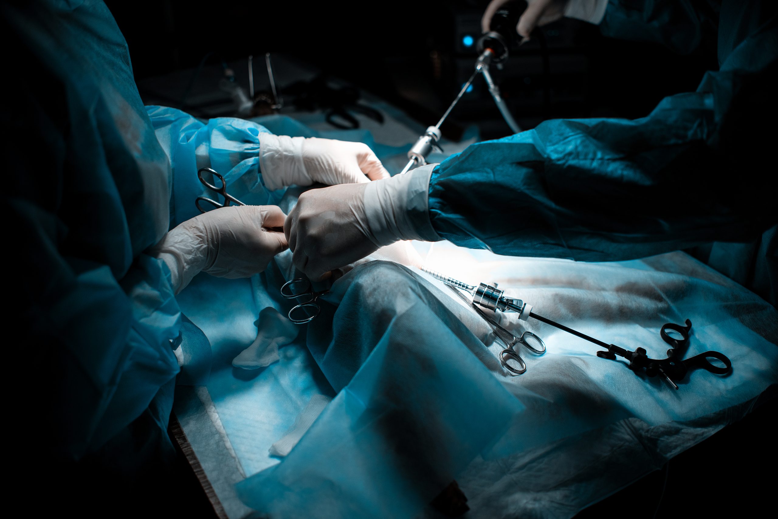
Procedure:
- Tube Selection:
- Various tracheostomy tubes are available, including cuffed and uncuffed options with or without an inner cannula.
- The choice depends on individual patient factors.
- Uncuffed tubes or deflated cuffs are preferred to reduce tracheal damage risk.
- Removable inner cannulas allow effective tube maintenance.
- Without an inner cannula, the entire tube should be replaced every 24 hours to prevent occlusion by secretions.
- Surgical Placement:
- A tracheostomy tube is placed surgically between the tracheal rings.
- It bypasses the upper airway, allowing oxygen flow distal to obstructions.
- The procedure is lifesaving and essential for patients with upper airway emergencies.
- Postoperative Care:
- Constant monitoring is crucial while the tube is in place.
- Complications (e.g., bleeding, airway damage, laryngeal paralysis) must be promptly recognized and treated.
- Emergency measures (such as tracheostomy tie or temporary neck hole for breathing) may be necessary.
- Some patients may require immediate intervention if complications arise.
Benefits:
- Airway Protection:
- Tracheostomy ensures a patent airway, especially when upper airway obstruction is critical.
- Long-Term Ventilation:
- It facilitates long-term positive pressure ventilation in critically ill patients.
- Reduced Oral Cavity Damage:
- By accessing the trachea distal to the mouth and pharynx, tracheostomy minimizes the risk of oral cavity damage during extended intubation.
Remember that individual cases may vary, and consulting a veterinarian is essential for proper diagnosis and treatment.